
More Information
Submitted: July 14, 2025 | Approved: July 24, 2025 | Published: July 25, 2025
How to cite this article: Yan L, Yongjing S, Shujuan H, Gong J, Tao X. The Role of Extracellular Vesicles in Synovial Injuries of Athletic Joints. J Sports Med Ther. 2025; 10(3): 035-045. Available from:
https://dx.doi.org/10.29328/journal.jsmt.1001095
DOI: 10.29328/journal.jsmt.1001095
Copyright License: © 2025 Yan L, et al. This is an open access article distributed under the Creative Commons Attribution License, which permits unrestricted use, distribution, and reproduction in any medium, provided the original work is properly cited.
Keywords: Extracellular vesicles; Synovial membrane injury; Inflammation regulation

The Role of Extracellular Vesicles in Synovial Injuries of Athletic Joints
Luo Yan, Song Yongjing, Hu Shujuan, Jianming Gong and Xiong Tao*
College of Education and Sports Sciences, Yangtze University, Jingzhou 423023, Hubei Province, China
*Address for Correspondence: Xiong Tao, College of Education and Sports Sciences, Yangtze University, Jingzhou 423023, Hubei Province, China, Email: [email protected]
Synovial membrane injury, which profoundly affects joint structure and function, plays a pivotal role in the progression of joint diseases. When manifest clinically as pain, inflammation, joint stiffness, or function impairment, such injuries may eventually advance to degenerative changes, cartilage damage, or arthritis, which significantly diminishes patients’ quality of life. In the field of joint regenerative medicine, particularly concerning sports-related synovial membrane injuries, extracellular vesicles (EVs) released from damaged synovial cells have emerged as a key research focus. These vesicles not only serve as indicators of synovial damage and inflammation, but may also be integral to the underlying pathophysiological processes of these injuries. EVs can influence crucial biological processes such as inflammatory response, cell proliferation, and fibrosis. Additionally, bioactive molecules within these vesicles, i.e., microRNAs, proteins, and metabolites, are intensively involved in the recovery and repair processes of joint injuries. Thus, a comprehensive understanding of their roles and mechanisms is essential for devising innovative therapeutic strategies and improving patient outcomes. This review seeks to elucidate the function of extracellular vesicles in synovial membrane injuries associated with sports and their potential clinical applications, aiming to advance 15 therapeutic approaches and enhance the management of joint diseases.
Synovial membrane injury refers to damage of the synovial tissue within the joint, which can be caused by factors such as trauma, overuse, degenerative changes, infections, and autoimmune diseases [1,2]. The consequences of these injuries are multifaceted, including persistent pain, joint swelling, effusion, functional impairment, and structural damage to the joint [3]. These issues can lead to an increased risk of secondary infections and systemic complications, which significantly decline the patient’s quality of life [4]. Prolonged inflammation and injury can result in cartilage degradation, bone destruction, and joint deformity, potentially culminating in degenerative conditions such as osteoarthritis [5]. In addition, synovial injury is also associated with rheumatoid arthritis, and the inflammatory response may also give rise to other complications, including cardiovascular and pulmonary diseases, thereby exacerbating the patient’s overall health risks [6,7].
Current treatment modalities for synovial joint injuries encompass pharmacotherapy (such as nonsteroidal anti-inflammatory drugs, corticosteroids, and immunosuppressants), physical therapy, surgical interventions, and injection therapies [8]. However, these approaches have notable limitations, including inadequate symptom relief, uncertain long-term efficacy, potential side effects, surgical risks, and variability in patient responses [9]. Despite their benefits in alleviating symptoms associated with synovial membrane injury, there remains a pressing need to develop novel therapeutic strategies to enhance treatment outcomes and improve patients’ quality of life.
The application of extracellular vehicles (EVs) in the context of synovial membrane injury represents a promising and rapidly evolving area of research [10]. EVs are small membrane-bound vesicles released by cells that facilitate intercellular communication and regulate a range of physiological and pathological processes [11]. Studies have shown that EVs derived from mesenchymal stem cells (MSCs) can mitigate the inflammatory response in synovial membrane cells by delivering anti-inflammatory factors and microRNAs (miRNAs) that suppress inflammation [12]. Additionally, the growth factors and miRNAs released from EVs can promote the proliferation and migration of synovial and chondrocyte cells, thereby accelerating tissue repair and facilitating cartilage regeneration [13].
Furthermore, EVs have the potential to modulate immune cell function, balancing pro-inflammatory and anti-inflammatory responses, and reducing immune-mediated synovial damage. They can also inhibit autoimmune responses by delivering factors associated with regulatory T cells (Tregs) [14]. In addition to their immunomodulatory effects, EVs can serve as natural drug delivery vehicles, especially targeting synovial tissue to deliver therapeutic agents or gene therapy payloads, thus enhancing treatment efficacy and minimizing side effects [15].
Basic concepts and structural characteristics of extracellular vesicles
Extracellular vehicles (EVs) are a class of small membrane vesicles released from cells to the outside of the cell, and they play important roles in a variety of biological processes, including intercellular communication, signal transduction, waste discharge, and viral propagation [16]. EVs can carry a variety of biologically active molecules, such as proteins, nucleic acids, lipids, and metabolites, which are capable of triggering specific biological responses within the recipient cell [17]. EVs can be classified into three subgroups based on their origin, size, and biological function: exo-vesicles, microvesicles, and exo-like vesicles. The outer vesicles are produced by the endoplasmic reticulum with a diameter of about 30-150 nm and carry abundant information molecules. The microvesicles have a diameter of 100-1000 nm and are released from the surface of the cell membrane. And the exocytosis-like vesicles are produced by apoptotic cells and have a diameter of 50-5,000 nm. EVs share the same membrane orientation as cells: they have extracellular domains of lipids and transmembrane proteins on their surfaces, encasing cytoplasmic components, including proteins and nucleic acids. Because EVs contain components from the original cell, EVs have also been used as a source of circulating biomarkers in biofluids. Recently, several comparative proteomics studies have identified a range of proteins that have been identified as specific for EV subtypes. However, specific EV subtype marker proteins have not been identified due to the difficulty in finding consistent markers across studies. EVs are thought to be a means for secretory cells to dispose of harmful or unwanted intracellular components, as well as an important mediator of communication with other cells. EVs carry a wide variety of substances that can transmit signals to induce physiological changes in recipient cells. For example, ligands on the surface of EVs can bind to cognate receptors on target cells, thereby mediating signaling cascades. Internalized EVs can be degraded and become a source of nutrients for the recipient cell. Substances in the EVs can also be transferred to the cytoplasm of the target cell and present activity in the cytoplasm. The mechanism of transfer of nonviral EVs is unknown. EVs can mediate the interaction of secretory cells with the surrounding extracellular matrix (ECM). In addition, EVs deposited in the ECM can also serve as indirect communication with neighboring cells. EVs also function as telecommunicators because they can be released into blood vessels or lymphatic vessels and deliver the substances they contain to distant target cells [18-20].
In addition, EVs play multiple functions in biological processes. They act as mediators of intercellular information transfer, facilitate intercellular communication and signalling, and participate in the regulation of basic cellular biological processes, such as proliferation, differentiation, migration, and apoptosis [21-23]. In addition, EVs play a key role in the immune response, regulating the activation, differentiation and function of immune cells, and influencing the strength and direction of the immune response [24].
They also respond to cellular stress and metabolic demands, maintain cellular homeostasis, and play a regulatory role in the interaction of cells with their microenvironment, such as cell adhesion, migration, and tissue repair [25]. Thus, EVs have important biological functions in cellular communication, immune regulation, and biological system complexity (Figure 1).
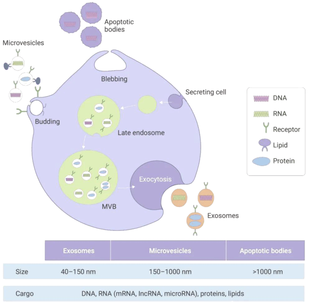
Figure 1: The formation and biological characteristics of EVs.
Overview of the pathophysiological mechanisms of synovial joint injuries
The synovium (SM) is a specialized loose connective tissue that lines the inner surface of the joint capsule [26]. It consists of two layers: the synovial membrane and the sub-synovial layer. The synovial membrane primarily contains macrophage-like synovial cells (MLS), fibroblast-like synovial cells (FLS), and a small number of dendritic-like synovial cells (DLS) [27,28]. The sub-synovial layer contains scattered blood vessels, lymphatic vessels, nerve fibers, adipocytes, fibroblasts, and a few lymphocytes or macrophages. Synovial cells produce lubricin and hyaluronic acid, which are components of the synovial fluid. The synovium functions as a semipermeable membrane, regulating the movement of molecules into and out of the joint cavity, maintaining the composition of synovial fluid, and lubricating the joint surface to reduce friction. Additionally, it provides nutrients to the avascular articular cartilage, helping to maintain its normal physiological state [29-31].
The synovial membrane is the inner lining of the joint, with primary functions including the secretion of Synovial fluid lubricates the joint, reduces friction, provides nutrients, and removes waste products.
Synovial membrane damage is common in various joint diseases, such as rheumatoid arthritis, osteoarthritis, and traumatic arthritis [32-34]. This damage is typically associated with an inflammatory response. Synovial cells and surrounding immune cells (such as macrophages and T cells) become activated, releasing various inflammatory mediators, including cytokines, chemokines, and prostaglandins. These mediators cause swelling, pain, and functional impairment of the synovial tissue [35].
Following joint injury, synovial cells may undergo abnormal proliferation, leading to thickening of the synovial tissue. This thickening can further compress joint structures and exacerbate the inflammatory response [36]. Inflammatory mediators can stimulate angiogenesis within the synovium, increasing local blood flow [37]. With this aid in repair, it may also lead to further infiltration of inflammatory cells and intensify the inflammatory response. Enzymes released during inflammation, such as matrix metalloproteinases and elastases, can degrade the matrix of the synovium and articular cartilage, leading to tissue destruction and loss of function [38]. In the context of chronic inflammation, synovial tissue may undergo fibrosis, characterized by the abnormal accumulation of collagen fibers. This fibrosis can result in joint stiffness and limited range of motion [39].
The synovial fluid, which is secreted by synovial cells within the joint cavity, has a specific composition and quantity under normal physiological conditions. However, when synovial damage occurs, this delicate balance may be disrupted [40]. In cases of inflammation, for instance, the body’s immune response is activated, leading to an increase in the production and release of inflammatory mediators.
These mediators, along with cellular debris from damaged cells, accumulate in the synovial fluid. As a Result, the normal physical and chemical properties of the synovial fluid are altered, further impacting the joints’ lubrication and cushioning functions that are crucial for smooth joint movement [35].
Interestingly, EVs have emerged as key players in this context. EVs can be released by various cell types within the joint environment, including synovial cells themselves. These EVs influence the function of synovial cells through various mechanisms. They can carry bioactive molecules such as proteins, nucleic acids, and lipids, which can be transferred to synovial cells upon interaction. In the healthy state, EVs may participate in maintaining the normal metabolism and function of synovial cells. However, in disease states, the content and function of EVs can change significantly. For example, in inflamed synovial membranes, EVs may carry pro - pro-inflammatory signals that exacerbate the inflammatory response or modulate the behavior of synovial cells in ways that contribute to joint damage. Thus, EVs play a complex and important role in both the health and disease states of the synovial membrane [41] (Figure 2).
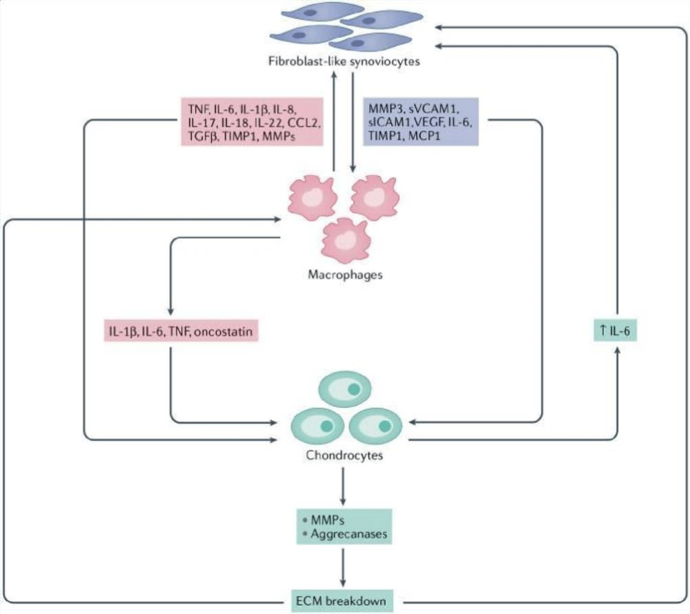
Figure 2: Cellular crosstalk in synovitis [42].
Generation and release of extracellular vesicles in synovial membranes
The biogenesis of extracellular vesicles consists mainly of the double invasion of the endoplasmic layer and the formation of intracellular multivesicular bodies (MVBs) containing intraluminal vesicles (ILVs). The ILVs are ultimately produced by exocytosis, which may be sized to be approximately 40 to 160 nm in diameter, by the plasma membrane and cytosolic spit, bound by the MVBs, which is then secreted out of the ILVs, whereas the first invasion of the plasmic layer produces a cup-shaped structure containing cell surface proteins and an extracellular environment, which is the ab initio formation of early sorting endosomes (ESEs), which can mature into late sorting endosomes (LSEs) and ultimately give rise to MVBs, also known as multivesicular endosomes [43]. Recently, researchers have found that EVs are key carriers of signaling molecules between joint and cartilage tissues, significantly contributing to the regulation of joint environment stability and influencing the pathogenesis of arthritis. Specifically, EVs are not only present in synovial fluid but are also widely distributed in articular cartilage, the synovial membrane, and the supernatant of chondrocytes, where they perform important biological functions [44].
In articular cartilage, EVs are considered active participants in intercellular interactions within tissues. They influence cartilage health by regulating extracellular matrix metabolism and inflammatory processes.
For example, exosomes secreted by synovial cells in response to IL-1β stimulation exhibit significantly different biological activities compared to exosomes secreted by unstimulated synovial cells. These IL-1β-stimulated exosomes significantly upregulate the expression of matrix metalloproteinase (MMP)-13 and ADAMTS-5 in chondrocytes while down-regulating the expression of key cartilage matrix proteins COL2A1 and ACAN, indicating their potential role in cartilage degeneration [45-47]. Additionally, ectosomes derived from IL-1β-stimulated synovial cells markedly enhance the migration and tube formation abilities of human umbilical vein endothelial cells, further inducing the release of more proteoglycans from cartilage explants. These changes are closely related to the development of joint degenerative diseases, particularly arthritis [48].
However, while inflammatory cytokines such as IL-6 and vascular endothelial growth factor (VEGF) have been detected in low levels in exosomes, more destructive inflammatory factors like IL-1β, tumor necrosis factor-α (TNF-α), matrix metalloproteinase-9 (MMP-9), and MMP-13 have not been detected in macrovesicles [49-51]. This suggests that different types of EVs may have significantly different roles in inflammatory responses. Recent research using nanostring analysis has shown that in IL-1β-stimulated synovial cells, there are significant differences in the expression levels of miRNAs in exosomes compared to unstimulated synovial cells. Notably, the expression levels of up to 50 miRNAs were significantly altered, indicating that these miRNAs may play important roles in the pathological processes of synovial cells and could potentially become targets for future therapeutic interventions [52,53].
Effect of extracellular vesicles on synoviocytes
EVs play multifaceted roles in synovial cells by transferring bioactive molecules, including the regulation of signaling pathways, inflammatory responses, cell proliferation and apoptosis, and matrix metabolism. Proteins, lipids, and RNA carried by EVs interact with synovial cells to modulate their functions, playing a crucial role in joint health and disease processes. Healthy synovial microenvironment primarily includes fibroblast-like synovial cells and macrophages [54,55]. Fibroblast-like synovial cells, derived from mesenchyme, are responsible for synthesizing key components of synovial fluid and extracellular matrix, such as lubricin and hyaluronic acid. The synovial lining, lacking a basement membrane, allows compounds to diffuse through its porous structure and may also facilitate the accumulation of immune triggers within the joint [56,57]. Wang, et al. discovered that miR-25-3p in EVs derived from fibroblast-like synovial cells can reduce chondrocyte necroptosis in knee osteoarthritis, alleviating disease severity [58]. Additionally, research indicates that in arthritis, EVs from fibroblast-like Synovial cells induce cellular senescence by down-regulating NF-κB and cAMP response element-related signaling pathways [28,59]. This process affects the production of pro-inflammatory factors and is closely related to the activation of pro-inflammatory phenotypes and the pathogenesis of chronic arthritis.
Macrophages in synovial tissue are classified into two subtypes: M1 and M2. Under physiological conditions, M2 are predominantly macrophages that maintain synovial tissue homeostasis. However, under certain environmental stimuli, macrophages can polarize into the M1 subtype, which produces pro-inflammatory cytokines and activates chondrocytes to secrete metalloproteinases [60]. This leads to cartilage degradation and promotes the development and progression of arthritis. Research has also shown that exosome-like microvesicles secreted by osteoarthritis chondrocytes can enhance IL-1β production in macrophages, exacerbating synovitis [53]. Zhang, et al. demonstrated that EVs derived from bone marrow mesenchymal stem cells can slow the progression of osteoarthritis, reduce cartilage damage, decrease osteophyte formation, inhibit M1 polarization of synovial macrophages, and promote M2 polarization [61].
In inflammatory joint diseases such as rheumatoid arthritis, EVs released by synoviocytes or immune cells can activate macrophages in the joints, causing them to secrete pro-inflammatory cytokines, leading to a vicious cycle of joint inflammation. These EVs also promote macrophage polarization to the M1 type, exacerbating damage to articular cartilage and synovium [62]. In addition, in the synovial tissue of osteoarthritis, large amounts of EVs released by inflammatory cells and synoviocytes activate macrophages and cause them to migrate to the site of inflammation. Activated macrophages secrete various inflammatory mediators such as prostaglandin E2 (PGE2) and MMPs, which can degrade collagen and proteoglycans in articular cartilage, destroying their structure and function. In addition, EVs can regulate macrophage polarization and promote its polarization to M1 type, which secretes inflammatory factors and reactive oxygen species (ROS) that can aggravate joint inflammation and cartilage damage [63]. This helps regulate synovial repair and regeneration, maintaining joint structural and functional stability (Figure 3).
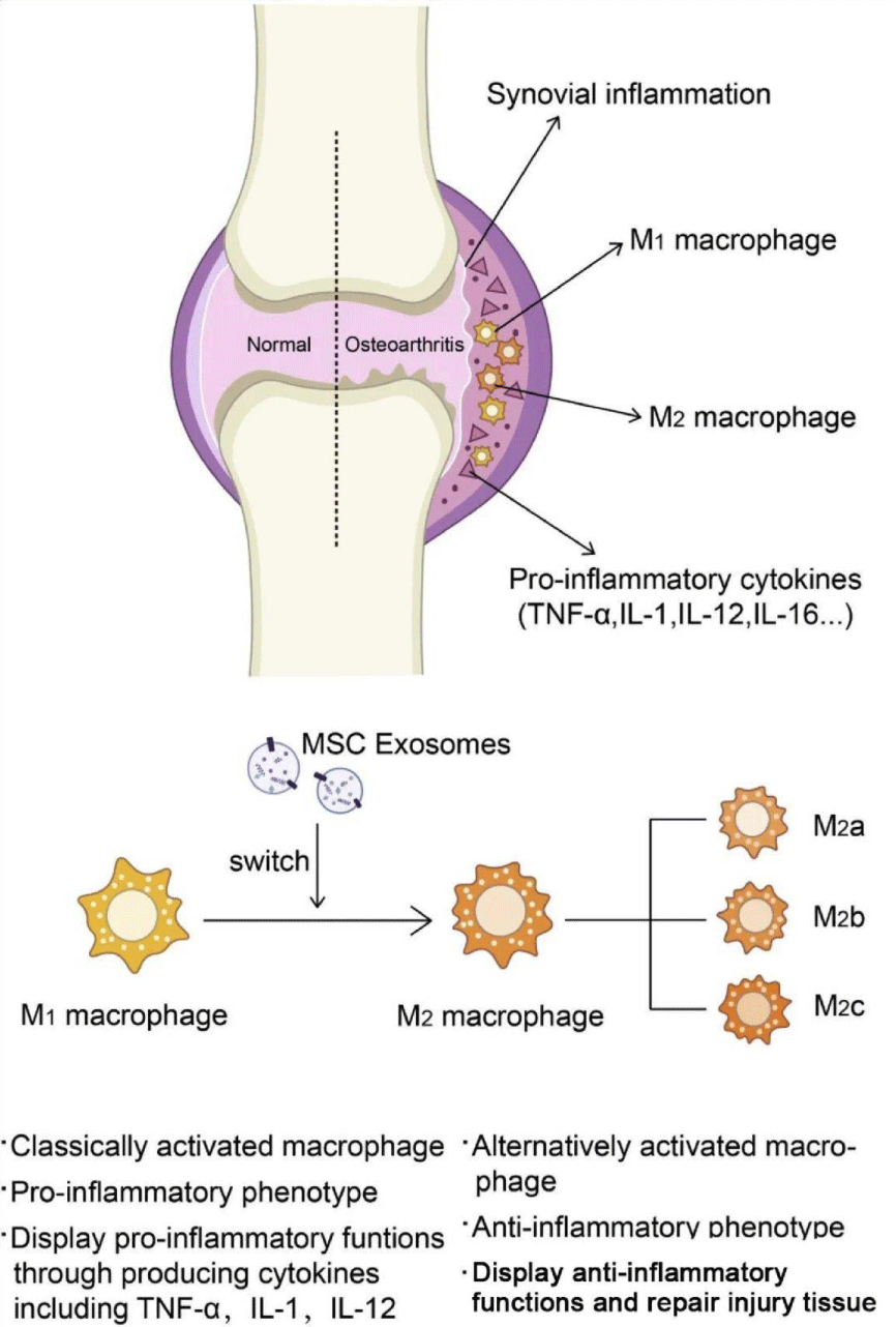
Figure 3: Macrophage activation and the role of extracellular vesicles of mesenchymal stem cells in synovial injury.
Role of extracellular vesicle-mediated signaling pathways in synovial damage
The NF-κB and PI3K/Akt signaling pathways are central to the onset and progression of synovial inflammation, serving as key drivers of injury and pathological changes. Their activation plays a pivotal role in shaping the inflammatory response within the synovium [64,65]. Research on EVs derived from synovial tissue indicates that they promote the expression of pro-inflammatory cytokines (such as IL-1β, IL-6, TNF-α, and COX-2) and cartilage degradation-related enzymes (such as MMP13, MMP9, and ADAMTS5) in chondrocytes, activating the NF-κB signaling pathway and leading to inflammation and degradation of cartilage [66-68]. Additionally, in synovial damage, activation of NF-κB and MAPK signaling pathways triggers the release of pro-inflammatory factors by synovial cells, contributing to the inflammatory response [69,70]. Under pathological conditions, EVs may carry molecules that can activate the NF-κB and MAPK signaling pathways, and these molecules enter synovial cells, triggering a signaling cascade that prompts synovial cells to release pro-inflammatory factors, such as TNF-α, IL-1β, and other pro-inflammatory factors, which exacerbate inflammatory responses. At the same time, EVs generated in the inflammatory environment may also carry more substances related to the activation of NF-κB and MAPK signaling pathways, forming a positive feedback mechanism that continues to amplify the inflammatory response, causing more serious damage to synovial tissue and joint function [71].
Mesenchymal stem cells (MSCs), known for their self-renewal and multi-lineage differentiation capabilities, are widely studied [72]. Among them, bone marrow-derived mesenchymal stem cells (BMSCs) play a critical role in bone remodeling [73]. Yu, et al. found that extracellular vesicles from BMSCs treated under hypoxic conditions may promote osteogenic repair in cranial osteoblasts via the PI3K/Akt signaling pathway [74]. Studies have also shown that TNF-α-induced activation of FLS in osteoarthritis activates the phosphorylation-dependent PI3K/Akt signaling pathway, increasing the expression of cadherin 11 in synovial fibroblasts, thereby aggravating synovitis and cartilage damage [75]. Thus, further investigation into the role of EVs in these signaling pathways is crucial for understanding the mechanisms of synovial-related diseases such as arthritis and for developing EV-based precision therapeutic strategies.
This not only provides new insights for future treatment options but also lays the foundation for personalized medicine.
Progress in the study of the correlation between extracellular vesicles and arthritis
EVs play a crucial role in the immune regulation of arthritis. They can carry antigen-presenting molecules, such as MHC class molecules, and interact with immune cells to modulate the activity of T cells, B cells, and macrophages, thereby influencing the immune system’s response [76,77]. Additionally, EVs are involved in the degradation of cartilage matrix in arthritis [78]. Research indicates that EVs can carry matrix metalloproteinases and members of the ADAMTS family, which are key players in cartilage degradation [79,80]. By promoting the expression and activity of these enzymes, EVs exacerbate cartilage damage and drive the progression of arthritis.
Due to the stability of EVs in body fluids and the diversity of their contents, they are considered potential diagnostic and prognostic biomarkers for arthritis [81]. Changes in EV miRNA derived from synovial fluid associated with joint alterations offer a unique opportunity to identify candidate biomarkers [82]. Studies have shown significant differences in the miRNA, protein profiles, and other molecular characteristics of EVs in patients with different types of arthritis. For instance, in rheumatoid arthritis serum EVs, miR-885-5p, miR-6894-3p, and miR-1268a are significantly upregulated in patient groups, suggesting potential as diagnostic markers [83]. In osteoarthritis patients, EVs from synovial fluid show varying miRNA profiles between genders: miR-16-2-3p is upregulated in female patients, while miR-26a-5p, miR-146a-5p, and miR-6821-5p are downregulated; in male patients, miR-6878-3p is downregulated while miR-210-5p is upregulated. In osteoblasts and osteoclasts, estrogen may regulate the expression and function of EV-related miRNAs. For example, estrogen may promote the increase of certain bone formation-associated miRNAs in EV secreted by osteoblasts, such as miR-21, which regulates signaling pathways related to osteoblast differentiation and promotes bone matrix synthesis [84].
These results also suggest that estrogen may play a significant role in EV-derived miRNAs [85]. Moreover, due to their inherent targeting capabilities and low immunogenicity, EVs are considered potential drug delivery carriers [86]. MSCs are of particular interest in arthritis treatment due to their repair and immunomodulatory capabilities [87]. MSC-derived EVs have been demonstrated to exert significant anti-inflammatory and chondroprotective effects. For example, hypoxia-treated MSC-EVs show enhanced cartilage repair abilities in animal models, possibly through promotion of osteoblast activity via PI3K/Akt signaling pathway [88]. Researchers are exploring gene engineering techniques to introduce anti-inflammatory miRNAs, proteins, or drug molecules into EVs for arthritis therapy. For instance, loading anti-inflammatory miRNAs (such as miR-223) into EVs has been shown to effectively suppress inflammation in arthritis in animal models [89]. In conclusion, further investigation into the mechanisms of EVs in the treatment of arthritis and other synovial-related diseases may provide new directions and insights for future precision therapies.
Extracellular vesicles as potential vehicles for the treatment of synovial injuries
Based on the important roles of EVs in synovial injuries, their potential as therapeutic targets have received intense attention in recent years. By modulating the production, release, or content of EVs, the pathological process of the synovium can be influenced, providing new therapeutic strategies [90]. It has been shown that bone marrow MSC-derived EVs prevent osteoarthritis by modulating synovial macrophage polarization, and in addition, synovial MSC-derived extracellular vesicles attenuate chondrocyte damage during osteoarthritis through microRNA-130b-3p-mediated inhibition of the LRP12/AKT/β-catenin axis [12,91]. Meng, et al. found that MSC-derived MicroRNA-320a in EVs regulate activation of rheumatoid arthritis fibroblast-like synoviocytes by inhibiting CXCL9 expression [92].
Although EVs are usually biocompatible, allogeneic EVs may trigger an immune response and their surface proteins or nucleic acid components may be immunogenic or toxic, increasing the safety risk [93]. Therefore, reducing the immunogenicity of EVs and ensuring their safety in humans are key challenges.
In addition, EVs have limited natural targeting ability and may be absorbed or rapidly cleared by non-targeted tissues after injection, compromising therapeutic efficacy [94]. Meanwhile, although EVs are capable of carrying a wide range of therapeutic molecules, how to efficiently load drugs and precisely control their release in target cells remain technical challenges, limiting their application in the treatment of complex diseases [95]. Therefore, the value of EVs in therapeutic applications can be significantly enhanced by engineering them to improve targeting, drug-carrying capacity, stability and reduce immunogenicity [96]. Such modification not only enhances the therapeutic efficacy of EVs, but also provides a powerful tool for the development of novel precision medicine [97]. Su, et al. constructed bone-targeted engineered probiotic EVs (BT-LGG-EVs) capable of delivering endogenous miRNAs to the bone microenvironment through the parabiotic bacterium Lactobacillus rhamnosus GG (LGG) for the targeted treatment of osteoporosis [98]. Zhang, et al. loaded chondrocyte-affinic peptide (CAP) onto the surface of the EVs using peptide-lipid coupling and loaded MMP13 siRNA using electroporation to construct CAP-Exo/siMMP13 with chondrocyte-targeting properties and attenuate cartilage degeneration by chondrocyte-specific silencing of MMP13 [99]. As a highly hydrophilic three-dimensional network material with good biocompatibility, adjustable mechanical properties, and efficient drug loading and release, hydrogels have been widely used in the fields of tissue engineering and drug delivery [100].
Osteogenesis-induced human dental pulp mesenchymal stem cells (DPSCs)-derived EV (Ost-EV) in combination with a multifunctional hydrogel effectively promoted bone tissue repair, and Ost-EV/hydrogel effectively alleviated inflammation, accelerated haemotransfusion, facilitated tissue calcification, and promoted bone tissue regeneration in an ectopic osteogenesis animal model [101]. Wu, et al. used a heat-sensitive hydrogel encapsulated with bone mesenchymal stem cell-derived sEVs by releasing exosome miR-21, accelerating osteogenesis and angiogenesis thereby treating synovial injury [102]. In addition, Yang, et al. developed an implantable hydroxyapatite (HAP)-embedded in situ crosslinked hyaluronic acid-alginate (HA-ALG) hydrogel system that combined EVs with a HAP-embedded in situ crosslinked HA-ALG hydrogel system to repair bone defects in rats in vivo [103]. In addition, targeted modification of EVs, using dual EVs combined with hydrogels, allows for better targeting and control of drug dose concentration and efficacy for better treatment of osteoarthritic diseases [104]. Extracellular vesicles exhibit significant therapeutic potential as vehicles for the treatment of synovial damage. However, technical, manufacturing and safety challenges need to be overcome to translate this potential into clinical applications. Future studies will continue to explore the use of EVs in synovial injuries and provide new therapeutic options for patients (Figure 4).
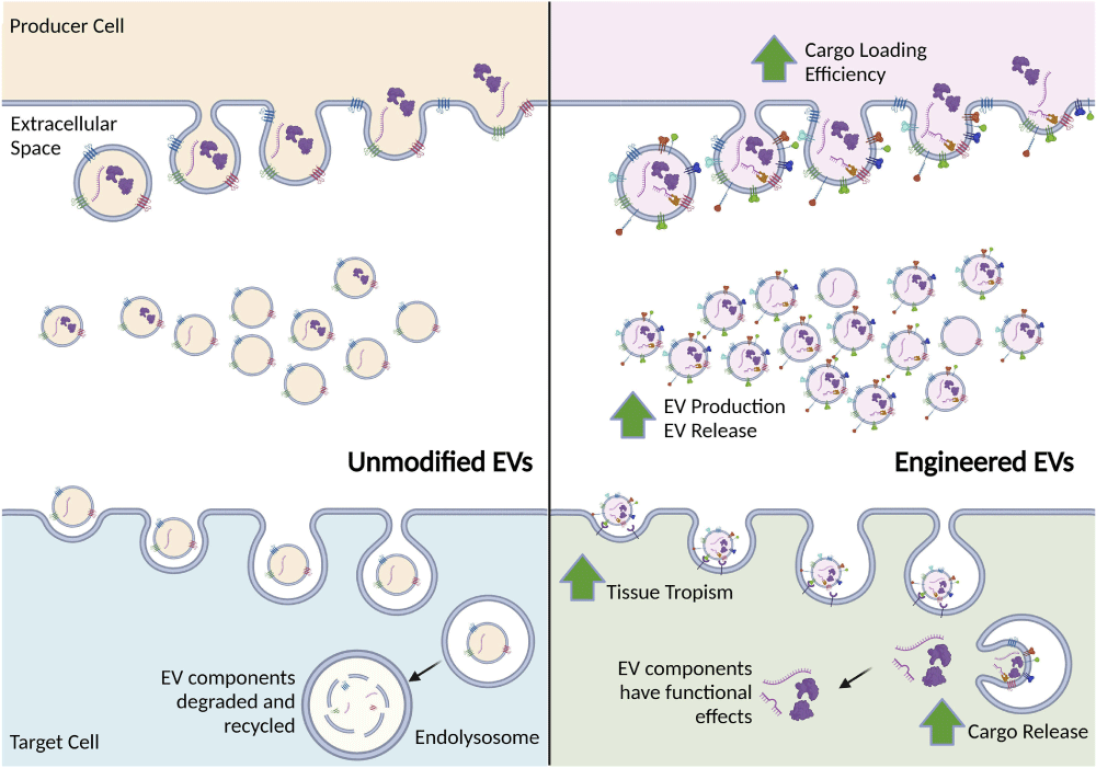
Figure 4: Endogenous and engineered EV pathways [105].
Future research directions and challenges
Although extensive research has revealed the role of EVs in synovial damage, the behind molecular mechanisms are still not fully understood. Future research could utilize multi-omics technologies (such as transcriptomics, proteomics, and metabolomics) to systematically analyze the content of EVs and their mechanisms of action in synovial cells to clarify these regulatory pathways [106-108]. Additionally, engineered EVs can be designed as highly targeted therapeutic carriers by incorporating functional nucleic acids or proteins [109]. Apart from major researches focus on improving the targeting, delivery efficiency, and therapeutic efficacy of EVs, particularly in applications related to chronic inflammatory diseases such as rheumatoid arthritis [110], another key research direction is to elucidate the dynamic distribution, clearance mechanisms, and biological effects of EVs in different tissues within the body. Future studies could use labeling and tracking technologies to pinpoint EVs’ action targets and their interactions with other biological systems [111].
Combining existing anti-inflammatory drugs and immune modulators with EV-based therapies to develop multimodal treatment approaches may offer more comprehensive therapeutic effects for synovial damage.
Researchers need to explore the synergistic effects of different treatment methods to optimize treatment regimens, enhance efficacy, and reduce side effects. However, despite the significant potential of EVs in basic research and clinical applications, the lack of standardized production and separation techniques remains a major challenge [112,113]. Future research should aim to achieve large-scale production and separation of high-purity, high-quality EVs without compromising their activity to meet clinical needs. Moreover, while EVs generally have good biocompatibility, their potential immunogenicity is a significant challenge. Particularly in the use of allogeneic EVs, reducing immune rejection and ensuring safety are key areas for future research [114].
Due to the heterogeneity of EVs and differences among patients and disease stages, their therapeutic effectiveness or sensitivity as diagnostic biomarkers may be affected [115]. Future research should investigate how the heterogeneity of EVs impacts their function and explore personalized treatment strategies. In summary, research on EVs in synovial damage is both promising and challenging. Future studies need to address key issues such as understanding the fundamental biological characteristics and pathological mechanisms of EVs, standardizing production, ensuring immunological safety, addressing individual differences, and translating findings to clinical applications. Through technological innovation and multidisciplinary collaboration, EVs have the potential to bring revolutionary breakthroughs in the diagnosis and treatment of synovial damage.
- Olivotto E, Belluzzi E, Pozzuoli A, Cigolotti A, Scioni M, Goldring SR, et al. Do synovial inflammation and meniscal degeneration impact clinical outcomes of patients undergoing arthroscopic partial meniscectomy? A histological study. Int J Mol Sci. 2022;23(7):3903. Available from: https://www.mdpi.com/1422-0067/23/7/3903
- Cao M, Ong MTY, Yung PSH, Tuan RS, Jiang Y. Role of synovial lymphatic function in osteoarthritis. Osteoarthritis Cartilage. 2022;30(9):1186-1197. Available from: https://www.sciencedirect.com/science/article/pii/S1063458422001261
- Lattanzi B, Lanni S, Luniew E, Giangiacomi M, Ravelli A, Jorini M. Ann Rheum Dis. 2015;74(Suppl 2):1221. Available from: https://www.proquest.com/docview/1901803606?sourcetype=Scholarly%20Journals#
- Pereira DF, Natour J, Buosi AL, Ferreira FB, Fernandes Ada R, Furtado RN. Is painless synovitis different from painful synovitis? A controlled, ultrasound, radiographic, clinical trial. Clinics (Sao Paulo). 2014;69(2):93-100. Available from: https://www.scielo.br/j/clin/a/y4c7vGhHrF6Kd8qTtjBdXYC/?lang=en
- Motta F, Barone E, Sica A, Selmi C. Inflammaging and osteoarthritis. Clin Rev Allergy Immunol. 2023;64(2):222-238. Available from: https://link.springer.com/article/10.1007/s12016-022-08941-1
- d'Ailly PN, Deugd C, Schep NWL, Kuijper TM, Kok MR, Willemze A, et al. Arthroscopic synovectomy versus intra-articular injection of corticosteroids for the management of refractory psoriatic or rheumatoid arthritis of the wrist: study protocol for a randomized controlled trial (ARCTIC trial). Trials. 2023;24(1):229. Available from: https://trialsjournal.biomedcentral.com/articles/10.1186/s13063-023-07129-y
- Ferguson LD, Siebert S, McInnes IB, Sattar N. Cardiometabolic comorbidities in RA and PsA: lessons learned and future directions. Nat Rev Rheumatol. 2019;15(8):461-474. Available from: https://www.nature.com/articles/s41584-019-0256-0
- Hohmann E. Editorial commentary: more clinical trials should focus on primary prevention of osteoarthritis: disruptive thinkers are required. Arthroscopy. 2021;37(8):2640-2641. Available from: https://www.arthroscopyjournal.org/article/S0749-8063(21)00586-3/fulltext
- Luft FC. Osteoarthritis is what the people have. J Mol Med. 2015;93:819–821. Available from: https://link.springer.com/article/10.1007/s00109-015-1316-9
- Ben-Trad L, Matei CI, Sava MM, Filali S, Duclos ME, Berthier Y, et al. Synovial extracellular vesicles: structure and role in synovial fluid tribological performances. Int J Mol Sci. 2022;23:11998. Available from: https://www.mdpi.com/1422-0067/23/19/11998
- Leidal AM, Debnath J. Unraveling the mechanisms that specify molecules for secretion in extracellular vesicles. Methods. 2020;177:15-26. Available from: https://www.sciencedirect.com/science/article/pii/S1046202320300213
- Harrell CR, Jovicic N, Djonov V, Arsenijevic N, Volarevic V. Mesenchymal stem cell-derived exosomes and other extracellular vesicles as new remedies in the therapy of inflammatory diseases. Cells. 2019;8(12):1605. Available from: https://www.mdpi.com/2073-4409/8/12/1605
- Pan C, Huang W, Chen Q, Xu J, Yao G, Li B, et al. LncRNA Malat-1 from MSCs-derived extracellular vesicles suppresses inflammation and cartilage degradation in osteoarthritis. Front Bioeng Biotechnol. 2021;9:772002. Available from: https://www.frontiersin.org/articles/10.3389/fbioe.2021.772002/full
- Wu R, Fan X, Wang Y, Shen M, Zheng Y, Zhao S, et al. Mesenchymal stem cell-derived extracellular vesicles in liver immunity and therapy. Front Immunol. 2022;13:833878. Available from: https://www.frontiersin.org/articles/10.3389/fimmu.2022.833878/full
- Alcaraz MJ. Control of articular degeneration by extracellular vesicles from stem/stromal cells as a potential strategy for the treatment of osteoarthritis. Biochem Pharmacol. 2024;228:116226. Available from: https://www.sciencedirect.com/science/article/pii/S0006295224001407
- van Niel G, Carter DRF, Clayton A, Lambert DW, Raposo G, Vader P. Challenges and directions in studying cell-cell communication by extracellular vesicles. Nat Rev Mol Cell Biol. 2022;23(5):369-382. Available from: https://www.nature.com/articles/s41580-022-00460-3
- Pathan M, Fonseka P, Chitti SV, Kang T, Sanwlani R, Van Deun J, et al. Vesiclepedia 2019: a compendium of RNA, proteins, lipids and metabolites in extracellular vesicles. Nucleic Acids Res. 2019;47(D1):D516-D519. Available from: https://academic.oup.com/nar/article/47/D1/D516/5146197
- Zhang W, Huang P, Lin J, Zeng H. The role of extracellular vesicles in osteoporosis: a scoping review. Membranes (Basel). 2022;12(3):324. Available from: https://www.mdpi.com/2077-0375/12/3/324
- Park SH, Lee EK, Yim J, Lee MH, Lee E, Lee YS, et al. Exosomes: nomenclature, isolation, and biological roles in liver diseases. Biomol Ther (Seoul). 2023;31:253-263. Available from: https://www.biomolther.org/journal/view.html?volume=31&number=3&spage=253
- Amin S, Massoumi H, Tewari D, Roy A, Chaudhuri M, Jazayerli C, et al. Cell type-specific extracellular vesicles and their impact on health and disease. Int J Mol Sci. 2024;25:2730. Available from: https://www.mdpi.com/1422-0067/25/5/2730
- Chen J, Yu X, Zhang X. Advances on biological functions of exosomal non-coding RNAs in osteoarthritis. Cell Biochem Funct. 2022;40:49–59. Available from: https://onlinelibrary.wiley.com/doi/10.1002/cbf.3679
- Du S, Ling H, Guo Z, Cao Q, Song C. Roles of exosomal miRNA in vascular aging. Pharmacol Res. 2021;165:105278. Available from: https://www.sciencedirect.com/science/article/pii/S1043661820303654
- Logvina NA, Shender VO, Arapidi GP, Holina TD. A role of vesicular transduction of intercellular signals in cancer development. Russ J Bioorg Chem. 2018;44:129–139. Available from: https://link.springer.com/article/10.1134/S1068162018010120
- Mittal S, Gupta P, Chaluvally-Raghavan P, Pradeep S. Emerging role of extracellular vesicles in immune regulation and cancer progression. Cancers (Basel). 2020;12:3563. Available from: https://www.mdpi.com/2072-6694/12/12/3563
- Zhou Z, Tao Y, Zhao H, Wang Q. Adipose extracellular vesicles: messengers from and to macrophages in regulating immunometabolic homeostasis or disorders. Front Immunol. 2021;12:666344. Available from: https://www.frontiersin.org/articles/10.3389/fimmu.2021.666344/full
- Kyung Chang S, Gu Z, Brenner MB. Fibroblast-like synoviocytes in inflammatory arthritis pathology: the emerging role of cadherin-11. Immunol Rev. 2010;233:256–266. Available from: https://onlinelibrary.wiley.com/doi/10.1111/j.0105-2896.2009.00854.x
- Li N, Gao J, Mi L, Zhang G, Zhang L, Zhang N, et al. Synovial membrane mesenchymal stem cells: past life, current situation, and application in bone and joint diseases. Stem Cell Res Ther. 2020;11:381. Available from: https://stemcellres.biomedcentral.com/articles/10.1186/s13287-020-01885-3
- Køster D, Egedal JH, Lomholt S, Hvid M, Jakobsen MR, Müller-Ladner U, et al. Phenotypic and functional characterization of synovial fluid-derived fibroblast-like synoviocytes in rheumatoid arthritis. Sci Rep. 2021;11:22168. Available from: https://www.nature.com/articles/s41598-021-01692-7
- Levick JR, McDonald JN. Fluid movement across synovium in healthy joints: role of synovial fluid macromolecules. Ann Rheum Dis. 1995;54:417–423. Available from: https://ard.bmj.com/content/54/5/417
- Jimenez SA, Derk CT. Following the molecular pathways toward an understanding of the pathogenesis of systemic sclerosis. Ann Intern Med. 2004;140:37. Available from: https://pubmed.ncbi.nlm.nih.gov/14706971/
- Hudalla GA. Medical applications of glycomaterials. Adv Healthc Mater. 2022;11:2200096. Available from: https://onlinelibrary.wiley.com/doi/10.1002/adhm.202200096
- Sadique M, Shah SR, Sharma SK, Islam SMN. Effect of significant parameters on squeeze film characteristics in pathological synovial joints. Mathematics. 2023;11:1468. Available from: https://www.mdpi.com/2227-7390/11/6/1468
- Liu Q, Zhang Y. Biological clock perspective in rheumatoid arthritis. Inflammation. 2025;48(3):1028–1041. Available from: https://link.springer.com/article/10.1007/s10753-024-02120-4
- Neumann E, Junker S, Schett G, Frommer K, Müller-Ladner U. Adipokines in bone disease. Nat Rev Rheumatol. 2016;12:296–302. Available from: https://www.nature.com/articles/nrrheum.2016.49
- Al-Madol MA, Shaqura M, John T, Likar R, Ebied RS, Salih MM, et al. Prostanoid receptor subtypes and its endogenous ligands with processing enzymes within various types of inflammatory joint diseases. Mediators Inflamm. 2020;2020:4301072. Available from: https://www.hindawi.com/journals/mi/2020/4301072/
- Philpott HT, Birmingham TB, Fiset B, Walsh LA, Coleman MC, Séguin CA, et al. Tensile strain and altered synovial tissue metabolism in human knee osteoarthritis. Sci Rep. 2022;12:17367. Available from: https://www.nature.com/articles/s41598-022-22459-8
- Hardy J, Bertone AL, Muir WW. Local hemodynamics, permeability, and oxygen metabolism during acute inflammation of innervated or denervated isolated equine joints. Am J Vet Res. 1998;59:1307. Available from: https://pubmed.ncbi.nlm.nih.gov/9781467/
- Pérez-García S, Carrión M, Gutiérrez-Cañas I, Villanueva-Romero R, Castro D, Martínez C, et al. Profile of matrix-remodeling proteinases in osteoarthritis: impact of fibronectin. Cells. 2019;9:40. Available from: https://www.mdpi.com/2073-4409/9/1/40
- Tamai K, Hamada J, Nagase Y, Morishige M, Naito M, Asai H, et al. Frozen shoulder. An overview of pathology and biology with hopes to novel drug therapies. Mod Rheumatol. 2024;34:439–443. Available from: https://academic.oup.com/mr/article/34/3/439/7663085
- Liu F, Lu Y, Wang X, Sun S, Pan H, Wang M, et al. Identification of FOXO1 as a geroprotector in human synovium through single-nucleus transcriptomic profiling. Protein Cell. 2024;15:441–459. Available from: https://academic.oup.com/protein-cell/article/15/5/441/7670122
- Wei Q, Zhu X, Wang L, Zhang W, Yang X, Wei W. Extracellular matrix in synovium development, homeostasis and arthritis disease. Int Immunopharmacol. 2023;121:110453. Available from: https://www.sciencedirect.com/science/article/pii/S1567576923002296
- Sanchez-Lopez E, Coras R, Torres A, Lane NE, Guma M. Synovial inflammation in osteoarthritis progression. Nat Rev Rheumatol. 2022;18:258–275. Available from: https://www.nature.com/articles/s41584-022-00749-9
- Zhao Y, Li X, Zhang W, Yu L, Wang Y, Deng Z, et al. Trends in the biological functions and medical applications of extracellular vesicles and analogues. Acta Pharm Sin B. 2021;11:2114–2135. Available from: https://www.sciencedirect.com/science/article/pii/S2211383521000594
- Malda J, Boere J, Van De Lest CHA, Van Weeren PR, Wauben MHM. Extracellular vesicles—new tool for joint repair and regeneration. Nat Rev Rheumatol. 2016;12:243–249. Available from: https://www.nature.com/articles/nrrheum.2015.170
- Horváth E, Sólyom Á, Székely J, Nagy EE, Popoviciu H. Inflammatory and metabolic signaling interfaces of the hypertrophic and senescent chondrocyte phenotypes associated with osteoarthritis. Int J Mol Sci. 2023;24:16468. Available from: https://www.mdpi.com/1422-0067/24/22/16468
- Tang Z, Feng H, Chen X, Shao S, Li C. SNORC knockdown alleviates inflammation, autophagy defect and matrix degradation of chondrocytes in osteoarthritis development. Mol Cell Biochem. 2023. Available from: https://link.springer.com/article/10.1007/s11010-023-04842-9
- Denkovskij J, Bagdonas E, Kusleviciute I, Mackiewicz Z, Unguryte A, Porvaneckas N, et al. Paracrine potential of the human adipose tissue-derived stem cells to modulate balance between matrix metalloproteinases and their inhibitors in the osteoarthritic cartilage in vitro. Stem Cells Int. 2017;2017:9542702. Available from: https://www.hindawi.com/journals/sci/2017/9542702/
- Kato T, Miyaki S, Ishitobi H, Nakamura Y, Nakasa T, Lotz MK, et al. Exosomes from IL-1β stimulated synovial fibroblasts induce osteoarthritic changes in articular chondrocytes. Arthritis Res Ther. 2014;16:R163. Available from: https://arthritis-research.biomedcentral.com/articles/10.1186/ar4679
- Kovács B, Vajda E, Nagy EE. Regulatory effects and interactions of the Wnt and OPG-RANKL-RANK signaling at the bone-cartilage interface in osteoarthritis. Int J Mol Sci. 2019;20:4653. Available from: https://www.mdpi.com/1422-0067/20/18/4653
- Łęgosz P, Sarzyńska S, Pulik Ł, Kotrych D, Małdyk P. The complexity of molecular processes in osteoarthritis of the knee joint. Open Med (Wars). 2020;15:366–375. Available from: https://www.degruyter.com/document/doi/10.1515/med-2020-0402/html
- Gao K, Zhu W, Li H, Ma D, Liu W, Yu W, et al. Association between cytokines and exosomes in synovial fluid of individuals with knee osteoarthritis. Mod Rheumatol. 2020;30:758–764. Available from: https://www.tandfonline.com/doi/full/10.1080/14397595.2019.1651445
- Liu B, Xian Y, Chen X, Shi Y, Dong J, Yang L, et al. Inflammatory fibroblast-like synoviocyte-derived exosomes aggravate osteoarthritis via enhancing macrophage glycolysis. Adv Sci (Weinh). 2024;11:2307338. Available from: https://onlinelibrary.wiley.com/doi/10.1002/advs.202307338
- Ni Z, Kuang L, Chen H, Xie Y, Zhang B, Ouyang J, et al. The exosome-like vesicles from osteoarthritic chondrocytes enhanced mature IL-1β production of macrophages and aggravated synovitis in osteoarthritis. Cell Death Dis. 2019;10:522. Available from: https://www.nature.com/articles/s41419-019-1739-2
- Liu G, Kang G, Wang S, Huang Y, Cai Q. Extracellular vesicles: emerging players in plant defense against pathogens. Front Plant Sci. 2021;12:757925. Available from: https://www.frontiersin.org/articles/10.3389/fpls.2021.757925/full
- Sun W, Cui H, Xu T, Yue J, Liang J, You W, et al. RNA binding proteins in extracellular vesicles and their potential value for cancer diagnosis and treatment (Review). Int J Oncol. 2023;63:114. Available from: https://www.spandidos-publications.com/10.3892/ijo.2023.5562
- Cheng WJ, Yang HT, Chiang CC, Lai KH, Chen YL, Shih HL, et al. Deer velvet antler extracts exert anti-inflammatory and anti-arthritic effects on human rheumatoid arthritis fibroblast-like synoviocytes and distinct mouse arthritis. Am J Chin Med. 2022;50:1617–1643. Available from: https://www.worldscientific.com/doi/10.1142/S0192415X22500689
- Bai Z, Bartelo N, Aslam M, Murphy EA, Hale CR, Blachere NE, et al. Synovial fibroblast gene expression is associated with sensory nerve growth and pain in rheumatoid arthritis. Sci Transl Med. 2024;16:eadk3506. Available from: https://www.science.org/doi/10.1126/scitranslmed.adk3506
- Wang J, Sun T. Mir-25-3p in extracellular vesicles from fibroblast-like synoviocytes alleviates pyroptosis of chondrocytes in knee osteoarthritis. J Bioenerg Biomembr. 2023;55:365–380. Available from: https://link.springer.com/article/10.1007/s10863-023-09964-9
- Xia ZB, Meng FR, Fang YX, Wu X, Zhang CW, Liu Y, et al. Inhibition of NF-κB signaling pathway induces apoptosis and suppresses proliferation and angiogenesis of human fibroblast-like synovial cells in rheumatoid arthritis. Medicine (Baltimore). 2018;97:e10920. Available from: https://journals.lww.com/md-journal/Fulltext/2018/06150/Inhibition_of_NF_B_signaling_pathway_induces.18.aspx
- Zhang H, Cai D, Bai X. Macrophages regulate the progression of osteoarthritis. Osteoarthritis Cartilage. 2020;28:555–561. Available from: https://www.sciencedirect.com/science/article/pii/S1063458420300539
- Zhang J, Rong Y, Luo C, Cui W. Bone marrow mesenchymal stem cell-derived exosomes prevent osteoarthritis by regulating synovial macrophage polarization. Aging (Albany NY). 2020;12:25138–25152. Available from: https://www.aging-us.com/article/104110/text
- Kim H, Back JH, Han G, Lee SJ, Park YE, Gu MB, et al. Extracellular vesicle-guided in situ reprogramming of synovial macrophages for the treatment of rheumatoid arthritis. Biomaterials. 2022;286:121578. Available from: https://www.sciencedirect.com/science/article/pii/S0142961222003306
- You B, Zhou C, Yang Y. MSC-EVs alleviate osteoarthritis by regulating microenvironmental cells in the articular cavity and maintaining cartilage matrix homeostasis. Ageing Res Rev. 2023;85:101864. Available from: https://www.sciencedirect.com/science/article/pii/S1568163723001070
- Wang L, He C. Nrf2-mediated anti-inflammatory polarization of macrophages as therapeutic targets for osteoarthritis. Front Immunol. 2022;13:967193. Available from: https://www.frontiersin.org/articles/10.3389/fimmu.2022.967193/full
- Ding Y, Chen Q. Recent advances on signaling pathways and their inhibitors in spinal cord injury. Biomed Pharmacother. 2024;176:116938. Available from: https://www.sciencedirect.com/science/article/pii/S0753332224002043
- Chen P, Zhou J, Ruan A, Guan H, Xie J, Zeng L, et al. Synovial tissue-derived extracellular vesicles induce chondrocyte inflammation and degradation via NF-κB signalling pathway: an in vitro study. J Cell Mol Med. 2022;26(4):2038–2048. Available from: https://onlinelibrary.wiley.com/doi/full/10.1111/jcmm.17227
- Ren C, Jin J, Hu W, Chen Q, Yang J, Wu Y, et al. Betulin alleviates the inflammatory response in mouse chondrocytes and ameliorates osteoarthritis via AKT/Nrf2/HO-1/NF-κB axis. Front Pharmacol. 2021;12:754038. Available from: https://www.frontiersin.org/articles/10.3389/fphar.2021.754038/full
- Miao Y, Wu S, Gong Z, Chen Y, Xue F, Liu K, et al. SPARCL1 promotes chondrocytes extracellular matrix degradation and inflammation in osteoarthritis via TNF/NF-κB pathway. J Orthop Transl. 2024;46:116–128. Available from: https://www.sciencedirect.com/science/article/pii/S2214031X24000168
- Fan W, Xu Z, Liang S, Zuo S, Bian C, Gao X, et al. MLL3 inhibits apoptosis of rheumatoid arthritis fibroblast-like synoviocytes promote secretion of inflammatory factors by activating CCL2 and the NF-κB pathway. Inflammation. 2021;44(5):1803–1814. Available from: https://link.springer.com/article/10.1007/s10753-021-01459-2
- Cao C, Wu F, Niu X, Hu X, Cheng J, Zhang Y, et al. Cadherin-11 cooperates with inflammatory factors to promote the migration and invasion of fibroblast-like synoviocytes in pigmented villonodular synovitis. Theranostics. 2020;10(24):10573–10588. Available from: https://www.thno.org/v10p10573.htm
- Wei K, Korsunsky I, Marshall JL, Gao A, Watts GFM, Major T, et al. Notch signalling drives synovial fibroblast identity and arthritis pathology. Nature. 2020;582(7811):259–264. Available from: https://www.nature.com/articles/s41586-020-2222-z
- Lan T, Luo M, Wei X. Mesenchymal stem/stromal cells in cancer therapy. J Hematol Oncol. 2021;14:195. Available from: https://jhoonline.biomedcentral.com/articles/10.1186/s13045-021-01208-w
- Hu LW, Wang X, Jiang XQ, Xu LQ, Pan HY. in vivo and in vitro study of osteogenic potency of endothelin-1 on bone marrow-derived mesenchymal stem cells. Exp Cell Res. 2017;357(1):25–32. Available from: https://www.sciencedirect.com/science/article/pii/S0014482717301393
- Deng J, Wang X, Zhang W, Sun L, Han X, Tong X, et al. Versatile hypoxic extracellular vesicles laden in an injectable and bioactive hydrogel for accelerated bone regeneration. Adv Funct Mater. 2023;33(14):2211664. Available from: https://onlinelibrary.wiley.com/doi/full/10.1002/adfm.202211664
- Liu S, Cao C, Zhang Y, Liu G, Ren W, Ye Y, et al. PI3K/Akt inhibitor partly decreases TNF-α-induced activation of fibroblast-like synoviocytes in osteoarthritis. J Orthop Surg Res. 2019;14:425. Available from: https://josr-online.biomedcentral.com/articles/10.1186/s13018-019-1394-4
- Wen M, Li Y, Qin X, Qin B, Wang Q. Insight into cancer immunity: MHCs, immune cells and commensal microbiota. Cells. 2023;12(14):1882. Available from: https://www.mdpi.com/2073-4409/12/14/1882
- Hu XX, Wu Y, Zhang J, Wei W. T-cells interact with B cells, dendritic cells, and fibroblast-like synoviocytes as hub-like key cells in rheumatoid arthritis. Int Immunopharmacol. 2019;70:428–434. Available from: https://www.sciencedirect.com/science/article/pii/S1567576918324007
- Sanchez C, Zappia J, Dierckxsens Y, Delcour JP, Henrotin Y. Boswellia serrata extract and curcumin increases GDF15 production by human primary osteoarthritis chondrocytes: a new mechanism of action. Osteoarthritis Cartilage. 2020;28(S1):S120. Available from: https://www.oarsijournal.com/article/S1063-4584(20)30263-6/fulltext
- Shimoda M. Extracellular vesicle-associated MMPs: a modulator of the tissue microenvironment. In: Makowski GS, editor. Adv Clin Chem. Vol. 88. Amsterdam: Elsevier; 2019;35–66. Available from: https://www.sciencedirect.com/science/article/abs/pii/S006524231830046X
- Thuault S, Ghossoub R, David G, Zimmermann P. A journey on extracellular vesicles for matrix metalloproteinases: a mechanistic perspective. Front Cell Dev Biol. 2022;10:886381. Available from: https://www.frontiersin.org/articles/10.3389/fcell.2022.886381/full
- Lättekivi F, Guljavina I, Midekessa G, Viil J, Heath PR, Bæk R, et al. Profiling blood serum extracellular vesicles in plaque psoriasis and psoriatic arthritis patients reveals potential disease biomarkers. Int J Mol Sci. 2022;23(7):4005. Available from: https://www.mdpi.com/1422-0067/23/7/4005
- Poulsen TBG, Andersen JS, Kristiansen MK, Rasmusen S, Arent-Nielsen L, Nielsen CH, et al. AB1254 phenotyping of multiple biofluids for liquid biomarkers for diagnostics and personalized medicine of rheumatoid arthritis, spondyloarthritis and osteoarthritis. Ann Rheum Dis. 2020;79(Suppl 1):1918–1919. Available from: https://ard.bmj.com/content/79/Suppl_1/1918.2
- Gong J, Zhang X, Khan A, Liang J, Xiong T, Yang P, et al. Identification of serum exosomal miRNA biomarkers for diagnosis of rheumatoid arthritis. Int Immunopharmacol. 2024;129:111604. Available from: https://www.sciencedirect.com/science/article/pii/S1567576924000834
- Maeda Y, Farina NH, Matzelle MM, Fanning PJ, Lian JB, Gravallese EM. Synovium-derived microRNAs regulate bone pathways in rheumatoid arthritis. J Bone Miner Res. 2017;32(3):461–472. Available from: https://asbmr.onlinelibrary.wiley.com/doi/10.1002/jbmr.3005
- Kolhe R, Hunter M, Liu S, Jadeja RN, Pundkar C, Mondal AK, et al. Gender-specific differential expression of exosomal miRNA in synovial fluid of patients with osteoarthritis. Sci Rep. 2017;7:2029. Available from: https://www.nature.com/articles/s41598-017-01905-y
- Ortega A, Martinez-Arroyo O, Forner MJ, Cortes R. Exosomes as drug delivery systems: endogenous nanovehicles for treatment of systemic lupus erythematosus. Pharmaceutics. 2020;13(1):3. Available from: https://www.mdpi.com/1999-4923/13/1/3
- Ansboro S, Roelofs AJ, De Bari C. Mesenchymal stem cells for the management of rheumatoid arthritis: immune modulation, repair or both? Curr Opin Rheumatol. 2017;29(2):201–207. Available from: https://doi.org/10.1097/bor.0000000000000370
- Li S, Liu J, Liu S, Jiao W, Wang X. Chitosan oligosaccharides packaged into rat adipose mesenchymal stem cells-derived extracellular vesicles facilitating cartilage injury repair and alleviating osteoarthritis. J Nanobiotechnol. 2021;19:343. Available from: https://doi.org/10.1186/s12951-021-01086-x
- Liu W, Liu A, Li X, Sun Z, Sun Z, Liu Y, et al. Dual-engineered cartilage-targeting extracellular vesicles derived from mesenchymal stem cells enhance osteoarthritis treatment via miR-223/NLRP3/pyroptosis axis: toward a precision therapy. Bioact Mater. 2023;30:169–183. Available from: https://doi.org/10.1016/j.bioactmat.2023.06.012
- Zhou X, Cao H, Guo J, Yuan Y, Ni G. Effects of BMSC-derived EVs on bone metabolism. Pharmaceutics. 2022;14(5):1012. Available from: https://doi.org/10.3390/pharmaceutics14051012
- Zhang J, Rong Y, Luo C, Cui W. Bone marrow mesenchymal stem cell-derived exosomes prevent osteoarthritis by regulating synovial macrophage polarization. Aging. 2020;12(24):25138–25152. Available from: https://doi.org/10.18632/aging.104110
- Meng Q, Qiu B. Exosomal microRNA-320a derived from mesenchymal stem cells regulates rheumatoid arthritis fibroblast-like synoviocyte activation by suppressing CXCL9 expression. Front Physiol. 2020;11:441. Available from: https://doi.org/10.3389/fphys.2020.00441
- Li JK, Yang C, Su Y, Luo JC, Luo MH, Huang DL, et al. Mesenchymal stem cell-derived extracellular vesicles: a potential therapeutic strategy for acute kidney injury. Front Immunol. 2021;12:684496. Available from: https://doi.org/10.3389/fimmu.2021.684496
- You DG, Oh BH, Nguyen VQ, Lim GT, Um W, Jung JM, et al. Vitamin A-coupled stem cell-derived extracellular vesicles regulate the fibrotic cascade by targeting activated hepatic stellate cells in vivo. J Control Release. 2021;336:285–295. Available from: https://doi.org/10.1016/j.jconrel.2021.06.031
- Kalluri R, LeBleu VS. The biology, function, and biomedical applications of exosomes. Science. 2020;367(6478):eaau6977. Available from: https://doi.org/10.1126/science.aau6977
- Lu Y, Yang Y, Liu S, Ge S. Biomaterials constructed for MSC-derived extracellular vesicle loading and delivery—a promising method for tissue regeneration. Front Cell Dev Biol. 2022;10:898394. Available from: https://doi.org/10.3389/fcell.2022.898394
- Dong Y, Xia P, Xu X, Shen J, Ding Y, Jiang Y, et al. Targeted delivery of organic small-molecule photothermal materials with engineered extracellular vesicles for imaging-guided tumor photothermal therapy. J Nanobiotechnol. 2023;21:442. Available from: https://doi.org/10.1186/s12951-023-02133-5
- Liu H, Wu Y, Wang F, Wang S, Ji N, Wang M, et al. Bone-targeted engineered bacterial extracellular vesicles delivering miRNA to treat osteoporosis. Compos Part B Eng. 2023;267:111047. Available from: https://doi.org/10.1016/j.compositesb.2023.111047
- Zhang H, Yan W, Wang J, Xie S, Tao WA, Lee CW, et al. Surface functionalization of exosomes for chondrocyte-targeted siRNA delivery and cartilage regeneration. J Control Release. 2024;369:493–505. Available from: https://doi.org/10.1016/j.jconrel.2024.04.009
- Wu Y, Li X, Wang Y, Shi Y, Wang F, Lin G. Research progress on mechanical properties and wear resistance of cartilage repair hydrogel. Mater Des. 2022;216:110575. Available from: https://doi.org/10.1016/j.matdes.2022.110575
- Wang L, Wei X, He X, Xiao S, Shi Q, Chen P, et al. Osteoinductive dental pulp stem cell-derived extracellular vesicle-loaded multifunctional hydrogel for bone regeneration. ACS Nano. 2024;18(7):8777–8797. Available from: https://doi.org/10.1021/acsnano.3c11542
- Wu D, Qin H, Wang Z, Yu M, Liu Z, Peng H, et al. Bone mesenchymal stem cell-derived sEV-encapsulated thermosensitive hydrogels accelerate osteogenesis and angiogenesis by release of exosomal miR-21. Front Bioeng Biotechnol. 2022;9:829136. Available from: https://doi.org/10.3389/fbioe.2021.829136
- Yang S, Zhu B, Yin P, Zhao L, Wang Y, Fu Z, et al. Integration of human umbilical cord mesenchymal stem cells-derived exosomes with hydroxyapatite-embedded hyaluronic acid-alginate hydrogel for bone regeneration. ACS Biomater Sci Eng. 2020;6(3):1590–1602. Available from: https://doi.org/10.1021/acsbiomaterials.9b01363
- Li S, Zheng W, Deng W, Li Z, Yang J, Zhang H, et al. Logic-based strategy for spatiotemporal release of dual extracellular vesicles in osteoarthritis treatment. Adv Sci. 2024;11:2403227. Available from: https://doi.org/10.1002/advs.202403227
- Cecchin R, Troyer Z, Witwer K, Morris KV. Extracellular vesicles: the next generation in gene therapy delivery. Mol Ther. 2023;31(5):1225–1230. Available from: https://www.cell.com/molecular-therapy-family/molecular-therapy/fulltext/S1525-0016(23)00021-7
- Carr HL, Turner JD, Major T, Scheel-Toellner D, Filer A. New developments in transcriptomic analysis of synovial tissue. Front Med. 2020;7:21. Available from: https://doi.org/10.3389/fmed.2020.00021
- Chen D, Yu H, Zhang Y, Huang Y. Metabolomic analysis of extracellular vesicles from human synovial fluids. Microchem J. 2022;177:107257. Available from: https://doi.org/10.1016/j.microc.2022.107257
- Yang B, Li X, Fu C, Cai W, Meng B, Qu Y, et al. Extracellular vesicles in osteoarthritis of peripheral joint and temporomandibular joint. Front Endocrinol. 2023;14:1158744. Available from: https://doi.org/10.3389/fendo.2023.1158744
- Qian K, Fu W, Li T, Zhao J, Lei C, Hu S. The roles of small extracellular vesicles in cancer and immune regulation and translational potential in cancer therapy. J Exp Clin Cancer Res. 2022;41:286. Available from: https://jeccr.biomedcentral.com/articles/10.1186/s13046-022-02492-1
- Wang C, Xu M, Fan Q, Li C, Zhou X. Therapeutic potential of exosome-based personalized delivery platform in chronic inflammatory diseases. Asian J Pharm Sci. 2023;18:100772. Available from: https://doi.org/10.1016/j.ajps.2022.100772
- Liu XC, Zhang HJ, Xu R, Liu J, Xia HF, Xie QH, et al. A NIR-II quantum dot-assisted dual-color imaging strategy enables simultaneous tracking of two subtypes of extracellular vesicles in vivo. Chem Eng J. 2024;492:152242. Available from: https://doi.org/10.1016/j.cej.2024.152242
- Mohammadipoor A, Hershfield MR, Linsenbardt HR, Smith J, Mack J, Natesan S, et al. Biological function of extracellular vesicles (EVs): a review of the field. Mol Biol Rep. 2023;50(22):8639–8651. Available from: https://doi.org/10.1007/s11033-023-08624-w
- Zhang C, Yang X, Jiang T, Yan C, Xu X, Chen Z. Tissue-derived extracellular vesicles: isolation, purification, and multiple roles in normal and tumor tissues. Life Sci. 2023;321:121624. Available from: https://doi.org/10.1016/j.lfs.2023.121624
- Nguyen J, Fuhrmann G. Extracellular vesicles — a versatile biomaterial. Adv Healthc Mater. 2022;11:e2200192. Available from: https://doi.org/10.1002/adhm.202200192
- Frampton AE, Giovannetti E. Diagnosing pancreatic ductal adenocarcinoma using plasma extracellular vesicle RNA profiles. Gut. 2020;69(3):404–405. Available from: https://doi.org/10.1136/gutjnl-2019-319896