
More Information
Submitted: 08 August 2019 | Approved: 15 August 2019 | Published: 16 August 2019
How to cite this article: Fabunmi AA, Oladipupo SO. Comparison of selected lower limb biomechanical variables between university of ibadan sportsmen with and without patellofemoral pain syndrome. J Sports Med Ther. 2019; 4: 067-072.
DOI: 10.29328/journal.jsmt.1001046
Copyright License: © 2019 Fabunmi AA, et al. This is an open access article distributed under the Creative Commons Attribution License, which permits unrestricted use, distribution, and reproduction in any medium, provided the original work is properly cited.
Keywords: Patellofemoral pain syndrome; Hamstring tightness; Static quadriceps angle; Ankle pronation

Comparison of selected lower limb biomechanical variables between university of ibadan sportsmen with and without patellofemoral pain syndrome
Ayodeji Ayodele Fabunmi* and Samson Oluwaseyi Oladipupo
Physiotherapy Department, College of Medicine, University of Ibadan, Ibadan, Oyo State, Nigeria
*Address for Correspondence: Ayodeji Ayodele Fabunmi, PhD, Senior Lecturer & Honorary Consultant, Physiotherapy, Physiotherapy Department, College of Medicine, University of Ibadan, Ibadan, Oyo State, Nigeria, Tel: +234-703-235-2397; +234-708-181-3558; Email: [email protected];
website: http://www.com.ui.edu.ng/index.php/dr-a-a-fabunmi
Patellofemoral pain syndrome is common among athletes who participate in jumping, running and pivoting sports. The aim of this study was to compare selected lower limb biomechanical variables between University of Ibadan students (athletes) with and without patellofemoral pain syndrome.
The research design for this study was a case control survey and a purposive sampling technique was used to recruit participants. Two hundred and twenty two (191(85.8%) males and 31 (14.2%) females) sportsmen participated in this study. The participants’ age was between 20-29 years. Fourty sportsmen tested positive to Clarke’s test while 27 sportsmen tested positive to Eccentric step test. Measurements of static quadriceps angle, hamstring tightness and navicular height were taken for all participants.
Data were analyzed using descriptive statistics of mean, standard deviation, percentages and inferential statistics of Independent ‘t’ test.
The mean lower limb biomechanical variables of participants with patellofemoral pain syndrome were 13.18 ± 2.37°, 106.46 ± 16.11° and 1.21 ± 0.61 cm while those without were 13.65 ± 2.46°, 128.95 ± 25.36° and 1.03 ± 0.58 cm for static quadriceps angle, hamstring tightness and navicular height respectively. There was no significant difference (p > 0.05) in selected lower limb biomechanical variables between participants with and without patellofemoral pain syndrome.
In conclusion there was no significant difference in static quadriceps angle, hamstring tightness and ankle pronation between participants with and without patellofemoral pain syndrome. It was recommended that PFPS development is probably multifactorial with other functional disorders of the lower extremity apart from the selected variables.
Patellofemoral Pain Syndrome (PFPS) is the most common knee joint pathology among young age groups [1], and is referred to as pain in the retropatellar and peripatellar regions [2]. The most common symptoms in patients with PFPS is pain during and after physical activity, during bodyweight loading of the lower extremities as in walking up/down stairs and squatting, and in sitting with the knees flexed. The symptoms are usually of gradual onset, although sometimes they may be acute as when caused by trauma, unilateral or bilateral, usually described as achy, but may be sharp [3]. Athletes, sometimes report stiffness or pain on prolonged sitting with the knees flexed [4], knee giving way or buckling; the perceived instability being likely due to inhibitory effect of pain on the effective contraction of the quadriceps, and should be differentiated from instability originating from a patellar dislocation, subluxation or ligamentous injury of the knee [5].
Patellofemoral pain syndrome (PFPS) accounts for 25% of all knee injuries treated in sports medicine clinics [6]. It was reported that almost 25%–30% of all injuries seen in a sports medicine clinic [7] and up to 40% of clinical visits for knee problems [8] are attributed to PFPS. It tends to occur more in the 10-35 years old, showing with high level of activity [9]. It has been suggested that the patella is highly affected by its surrounding structures and has high instability, with the instability appearing to be higher in young adults with high level of activity than other age groups [10].
Patellofemoral pain syndrome is associated with various biomechanical characteristics of the lower extremity and potential risk factors [11]. Several risk factors play a part in the pathogenesis of PFPS with overuse, trauma and internal risk factors being particularly important among athletes [12]. Patellofemoral pain syndrome is associated with external and internal risk factors. The external risk factors are said to be found in sports activities or sports training habits, surrounding environments and incorrect use of sports equipments, while the internal risks are due to individual physical characteristics and psychological tendencies [13]. The internal risk factors can be anatomical or biomechanical (muscle tightness or weakness, generalized joint laxity, excessive ankle pronation, gait abnormalities, excessive quadriceps angle and so on) [14]. The biomechanical risk factors are explained in relation to anatomic location of the knee, which include proximal (upper femur, hip and trunk), local (in and around the patella and the patellofemoral joint) and distal (lower leg, foot and ankle) [14].
Patellofemoral pain syndrome is common among sports disciplines such as basketball [15], volleyball [16] and running [17]. Prevalence rates of between 13% and 26% are reported in females participating in soccer, volleyball, running, fencing and rock climbing [16]. It was reported that amateur athletes diagnosed with PFPS will stop participating in sports because of knee pain [18].
Clinical assessment and treatment of patellofemoral pain syndrome are extremely challenging because of the multiple forces affecting the patellofemoral joint. Wilk, et al. [19], opined that PFPS remains one of the most vexatious clinical challenges in rehabilitative medicine. Many patients with PFPS symptoms experience spontaneous recovery and even live without any symptoms at al. [20]. A combination of factors, such as abnormal lower limb biomechanics, soft-tissue tightness, muscle weakness, and excessive exercise, may result in increased cartilage and subchondral bone stress, and subtle patellar misalignment or more overt patellar maltracking. Fulkerson, [21] states that while opinions vary, there is believe that imbalance (misalignment) of the extensor mechanism can lead to overload of the retinaculum and subchondral bone. In particular, patellofemoral pain syndrome (PFPS), also known as anterior knee pain, makes up the significant portion of the overuse injuries observed in adolescent athletes [22]. Patellofemoral pain syndrome for a prolonged period in young adults tends to develop into degenerative arthritis of the patellofemoral joint, and symptoms of degenerative arthritis in old age can be predicted. Early diagnosis of PFPS is very important, and PFPS can be fully prevented before surgical intervention is needed via this early diagnosis [11]. The aim of this study was to compare selected lower limb Biomechanical variables between University of Ibadan Sportsmen with and without PFPS.
The research design for this study was a case control. Ethical approval was sought and obtained from the University of Ibadan/University College Hospital (UI/UCH) Health Research Ethics Committee. The rationale and procedure for the study were explained to the participants and their informed consent was obtained. Eccentric step test and Clarke’s test were used to evaluate University of Ibadan sportsmen to determine those with or without patellofemoral pain syndrome. A self-designed proforma was used to obtain/record socio-demographic data (gender, age, weight, height and types of sports) and lower limb biomechanical variables (static quadriceps angle, navicular height and knee extension angle) of each participant. Completed questionnaire was collected immediately or the following day where immediate collection was not possible.
Participants
Participants for this study were athletes whose sports of interest is one of football, running or basketball in University of Ibadan, without any form of remuneration. Participants with lower limb pathology (e.g. fracture-dislocation of the knee joint etc.) and with previous history of knee surgery were not allowed to participate in this study. Two hundred and twenty two sportsmen were randomly selected to participate in this study. One hundred and ninety one (85.8%) were males and 31 (14.2%) were females sportsmen. The participants age was between 20-29 years. Fourty sportsmen tested positive to Clarke’s test while 27 sportsmen tested positive to Eccentric step test.
Instruments
The following instruments were used for data collection:
Universal goniometer:
This device was used to measure the following:
a. Hamstrings Shortening: the angle of knee extension with the hip in 90° of flexion.
b. Static Quadriceps Angle: the angle between the quadriceps muscles and the patella tendon.
Ruler: A ruler with uniform markings in centimetres was used to measure the height of the navicular bone.
Height meter (SECA 220, Germany): This was calibrated in centimetres (from 20-210 cm). It was used to measure height of the participants to the nearest centimetres. Height was measured with participants standing with barefoot, with heads straight and back against the upright height meter. The perpendicular bar (lever arm of the height meter) was in contact with the vertex of the participant’s head with a light pressure applied on the participants’ heads. The height was then read off to the nearest centimeter.
Weighing scale (Hanson, Ireland): This was calibrated in kilogrammes (from 0-220 g). It was used to measure the participant’s body weight to the nearest 0.1 kg. The body weight of each participant was measured with a bathroom weighing scale. The participants were instructed to stand erect and bare-footed on the weighing scale with minimal clothing while looking ahead with hands held on either side of the body. The body weight was then read off to the nearest kilogram.
Proforma form: A self designed proforma was used to obtain data on athlete’s socio-demographic information such as gender, age, weight, height, types of sports and level of competition as well as athletes’ static quadriceps angle, navicular bone height and knee extension angle.
A step: A 15 cm tall stool/step was designed for the purpose of evaluation of participants for eccentric step test.
Evaluation tests
The following tests were used to screen participants for the presence of patellofemoral pain syndrome. Participants, who tested positive for either of the two tests, were classified as those with patellofemoral pa in syndrome while those who tested negative to both tests were classified as having no patellofemoral pain syndrome (control group). This is the gold standard as used in similar study by Kwon, et al. [23]. The tests included:
Eccentric step test: The participants performed the test with bare feet. The step was 15 cm high. Briefly, the participants were asked to stand on the step, put hands on the hips and step down from the step as slowly and smoothly as possible. Participants kept the hands on their hips throughout the test performance. After the participants have performed the test with one leg, the procedure was repeated using the other leg. The eccentric step test was considered positive for patellofemoral pain syndrome when the participant reported knee pain during the test performance [24].
Clarke’s test: Clarke’s test was performed with the participants lying supine with both knees supported by a pillow, in order to create an adequate amount of knee flexion (10°–20°) [25]. While the patient was relaxed, the examiner pressed the patella distally (with the hand on the superior border of the patella) and then asked the patient to contract the quadriceps muscle [26]. If the patient’s pain was reproduced during the test, the test was considered positive for patellofemoral pain syndrome.
Measurement of biomechanical variables
Assessment of hamstring flexibility: Participants’ hamstring flexibility was assessed using the hamstring flexibility test [27]. The test assessed the hamstring muscle length by measuring the angle of knee extension with the hip in 90° of flexion. Each participant performed the test twice on both lower limbs. Participants were positioned with the thigh supported by the plinth, the untested knee joint was put in 90° flexion across the edge of the plinth with the thigh of the untested leg resting on the plinth and stabilized with a strap to eliminate any elevation of the limb off the plinth and also the front of the participant’s pelvis was stabilized to maintain the pelvis in a neutral position during hamstring measurements. The participant was then asked to flex the hip of the tested to 90° and with the goniometer being used to ensure 90° hip flexion. With the axis of the goniometer placed over the greater trochanter, the stationary arm parallel to the midaxillary line of the trunk and the moveable arm parallel to the femur in line with the lateral femoral condyle [27]. The participant was then asked to straighten the knee joint as far as possible while maintaining the hip at 90 degrees. The axis of the goniometer was placed over the lateral knee joint line, the moveable arm aligned with the lateral malleolus of the ankle and the stationary arm aligned with the greater trochanter parallel to the femur [27]. The goniometer measured the angle of knee extension in degrees giving an indication of hamstring muscle length as shown in figure 1.
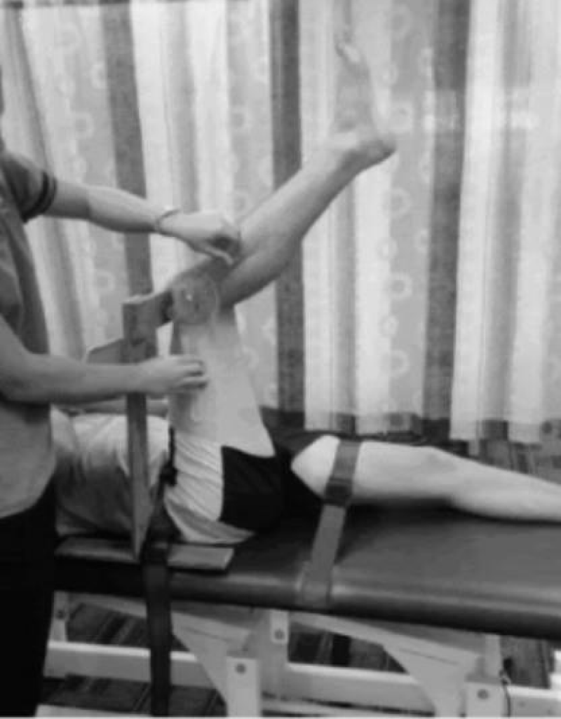
Figure 1: Assessment of Hamstring Flexibility.
Navicular drop test: The Navicular Drop Test (NDT) was first described by Brody, in 1982 as a means of quantifying the degree of ankle pronation in runners [28]. It was intended to represent the sagittal plane displacement of the navicular tuberosity from a neutral position to a relaxed position in standing [29]. Participants were in standing to ensure full weight-bearing through the lower extremity while the researcher ensured that the subtalar joint was in a neutral position. A mark was placed on the navicular tuberosity and its distance from the supporting surface (floor or step) measured (first measurement). The participant would then relax after the initial measurement while the researcher measured the amount of sagittal plane excursion of the navicular bone from the supporting surface with a ruler (second measurement) [30]. A difference of > 10 mm between the first and second measurements was considered significant excessive ankle pronation.
Static quadriceps angle (Q-angle): To measure the static Q-angle, the participants stood without shoes and their knees straight in a comfortable position while looking straight ahead. Anatomical landmarks including the border of the patella, the mid-patella, tibia tubercule and anterior superior iliac spine (ASIS) were palpated and marked. The axis of the goniometer was then placed on the midpoint of the patella, its stationary arm on the ASIS while the movable arm was aligned with the tibia tubercle. The static quadriceps angle was then measured as the angle between the line connecting the mid-patella and ASIS, and the line connecting the center of tibia tubercule and the patella [31].
Statistical analysis
Data was summarized using descriptive statistics of mean, standard deviation and percentages and inferential statistics of Independent t-test was used to compare the selected lower limb biomechanical variables between University of Ibadan sportsmen with and without patellofemoral pain syndrome. Level of significance (α) was set at 0.05.
Socio-demographic characteristics of participants.
A total of two hundred and fifty questionnaires were administered for the purpose of this study with two hundred and twenty-two questionnaires completely filled and returned. Two hundred and twenty-two University of Ibadan students (sportsmen) participated in this study; 40 sportsmen tested positive to Clarke’s test while 27 sportsmen tested positive to Eccentric step test. There were 191 male (86%) participants compared to 31 females (14%), while most of the participants fall within the age group of 20-29 years.
The mean and standard deviation of participants’ age, weight, height and body mass index (BMI) for sportsmen with and without PFPS for both test (Clarke’s test and Eccentric step test) are shown in table 1. There was a significant difference (p < 0.05) in height between participants with and without patellofemoral pain syndrome for both Clarke test and Eccentric step test (Table 1).
| Table 1: Independent t-test comparison of demographic variables between participants with and without patellofemoral pain syndrome for Eccentric step test and Clarke test | ||||||
| Variable | Clarke’s test | Eccentric step test | ||||
| Positive (N = 40) | Negative (N = 182) | p - value | Positive (N = 27) | Negative (N = 1 95) | p - value | |
| mean ± SD | mean ± SD | |||||
| Age (yrs): | 22.93 ± 3.17 | 21.95 ± 3.60 | 0.115 | 22.96 ± 3.47 | 22.01 ± 3.54 | 0.19 |
| Weight (kg): | 70.33 ± 10.47 | 68.97 ± 10.97 | 0.476 | 72.00 ± 13.04 | 68.83 ± 10.51 | 0.155 |
| Height (m): | 1.77 ± 0.07 | 1.73 ± 0.09 | 0.018* | 1.77 ± 0.07 | 1.73 ± 0.09 | 0.016* |
| BMI (kg/m²): | 22.59 ± 3.52 | 23.03 ± 3.03 | 0.422 | 22.83 ± 3.80 | 22.96 ± 3.02 | 0.835 |
| *indicates significant difference, p < 0.05 SD: Standard Deviation. |
||||||
Comparison of selected lower limb biomechanical variables between sportsmen with and without patellofemoral pain syndrome.
The mean and standard deviation values of lower limb biomechanical variables (static quadriceps angle, knee extension angle and navicular height) of University of Ibadan sportsmen with and without patellofemoral pain syndrome for both Clarke test and Eccentric step test were presented in table 2.
| Table 2: Independent t-test comparison of lower limb biomechanical variables between participants with and without patellofemoral pain syndrome using Eccentric step test and Clarke test. | ||||||
| Variable | Clarke | Eccentric | ||||
| Positive (N = 40) | Negative (N = 182) | p - value | Positive (N = 27) | Negative (N = 195) | p - value | |
| mean ± SD | mean ± SD | mean ± SD | mean ± SD | |||
| Static Q-angle (deg°): | 13.18 ± 2.37 | 13.65 ± 2.46 | 0.269* | 13.67 ± 2.88 | 13.55 ± 2.39 | 0.815* |
| Knee extension angle (deg°): | 128.95 ± 25.36 | 106.46 ± 16.11 | 0.064* | 106.07 ± 13.33 | 111.12 ± 15.30 | 0.724* |
| Navicular height (cm): | 1.21 ± 0.61 | 1.03 ± 0.58 | 0.080* | 1.22 ± 0.59 | 1.04 ± 0.59 | 0.139* |
| *indicates no significant difference, p > 0.05 SD: Standard Deviation. |
||||||
The mean and standard deviation values of lower limb biomechanical variables of University of Ibadan sportsmen with patellofemoral pain syndrome were 13.18 ± 2.37°, 128.95 ± 25.36° and 1.21 ± 0.61 cm for those who tested positive for Clarke test and 13.67 ± 2.88°, 106.07 ± 13.33° and 1.22 ± 0.59 cm for Eccentric step test for static quadriceps angle, knee extension angle and navicular height respectively (Table 2).
A comparison of the variables between University of Ibadan sportsmen with and without patellofemoral pain syndrome showed that there was no significant difference (P > 0.05) in mean values of lower limb biomechanical variables between sportsmen with and without patellofemoral pain syndrome for both Clarke test and Eccentric step test.
Three different sports were selected for the purpose of this study; football, basketball and running. The selection of these sports was based on the findings that patellofemoral pain syndrome is one of the most common overuse injuries among different sports disciplines such as basketball [15], volleyball [16] and running [17].
A higher proportion of participants with and without patellofemoral pain syndrome in this study, were within 20-29 years age group. This agrees with Baker, et al. [1] that patellofemoral pain syndrome is the most common knee joint pathology among young age groups, corroborated by Muller and Snyder-Mackler, [9] that patellofemoral pain syndrome tends to occur more in the 10-35 years of age, showing with high level of activity. There were one hundred and ninety-one (191) males (86%) who participated in the study compared to 31 females (14%) with a higher proportion of male participants with patellofemoral pain syndrome.
Elevated Q angle seems to be one of the suggested factors contributing to PFPS [32]. It has been theorized that an excessive static Q-angle is related to development of patellofemoral pain syndrome, through increased compressive forces between the lateral facet of the patella and the lateral femoral condyle [33]. The outcome of this study showed there was no significant difference in static Q angle measured between sportsmen with and without patellofemoral syndrome which is in agreement with Silva, et al. [32]. Silva, et al. [32], also compared dynamic Q angle between sportsmen with and without PFPS and obtained a significant difference between sportsmen with and without PFPS and hence were of the opinion that the dynamic Q angle measure is more predictive of PFPS than static Q angle measure. Thijs, et al. [34], and Ramskov, et al. [35], prospectively monitored amateur street runners and found that there is no significant difference in static Q-angle between athletes with and without patellofemoral pain syndrome and this is in agreement with result of this study.
Patil, et al. [36] identified a significant difference in hamstring tightness in patients with patellofemoral pain syndrome relative to the control group, this is contrary to the findings of this study in which there was no significant difference in hamstring tightness between University of Ibadan sportsmen with and without patellofemoral pain syndrome for both Clarke test and Eccentric step test. White, et al. [37], observed that sportsmen with PFPS presents with significant higher hamstring tightness compared with sportsmen without PFPS, which is not in agreement with results of this study.
Barton, et al. [38] showed that patients with patellofemoral pain syndrome have more pronated ankle type, increased forefoot abduction and increased rear-foot eversion in comparison with a healthy control group. Mølsgaard, et al. [39], also demonstrated abnormalities of the navicular bone in high school students with patellofemoral pain syndrome. Contrary to all the report of all these findings, there was no significance difference in navicular bone height (ankle pronation) between University of Ibadan sportsmen with and without patellofemoral pain syndrome (for both Clarke test and Eccentric step test) based on the findings of this study.
In conclusion there was no significant difference in selected lower limb biomechanical variables (static quadriceps angle, hamstring tightness and navicular height) between University of Ibadan sportsmen with and without patellofemoral pain syndrome. It was recommended that PFPS development is probably multifactorial with other functional disorders of the lower extremity apart from the selected variables in this study.
- Baker V, Bennell K, Stillman B, Cowan S, Crossley K. Abnormal knee joint position sense in individuals with patellofemoral pain syndrome. J Orthop Res. 2002; 20: 208-214. PubMed: https://www.ncbi.nlm.nih.gov/pubmed/11918299
- Loudon JK, Gajewski B, Goist-Foley HL, Loudon KL. The effectiveness of exercise in treating patellofemoral-pain syndrome. J Sport Rehabil. 2004; 13: 323-342.
- Hart H, Ackland D, Pandy M, Crossley K. Quadriceps volumes are reduced in people with patellofemoral joint osteoarthritis. Osteoarthritis Cartilage. 2012; 20: 863-868. PubMed: https://www.ncbi.nlm.nih.gov/pubmed/22525223
- Dixit S, DiFiori JP, Burton M, Mines B. Management of patellofemoral pain syndrome. Am Fam Physician. 2007; 15: 75: 94-202. PubMed: https://www.ncbi.nlm.nih.gov/pubmed/17263214
- Collado H, Fredericson M. Patellofemoral pain syndrome. Clin Sports Med. 2010; 29: 379-398.
- Brukner P, Khan K, McConnell J, Cook J. Anterior knee pain. In: Brukner P, Khan K. Clinical Sports Medicine. 2nd edn. New York, N.Y: McGraw-Hill RAW: 454-493.
- Witvrouw E, Callaghan MJ, Stefanik JJ, Noehren B, Bazett-Jones DM, et al. Patellofemoral pain: consensus statement from the 3rd International Patellofemoral Pain Research Retreat held in Vancouver, September 2013. Br J Sports Med. 2014; 48: 411-414. PubMed: https://www.ncbi.nlm.nih.gov/pubmed/24569145
- Natri A, Kannus P, Järvinen M. Which factors predict the long-term outcome in chronic patellofemoral pain syndrome? A 7-yr prospective follow-up study. Med Sci Sports Exerc. 1998; 30:1572-1577. PubMed: https://www.ncbi.nlm.nih.gov/pubmed/9813868
- Muller K, Snyder-Mackler L. Diagnosis of patellofemoral pain after arthroscopic meniscectomy. J Orthop Sports Phys Ther. 2000; 30: 138-142. PubMed: https://www.ncbi.nlm.nih.gov/pubmed/10721509
- Hamstra-Wright KL, Swanik CB, Ennis TY, Swanik KA. Joint stiffness and pain in individuals with patellofemoral syndrome. J Orthop Sports Phys Ther. 2005; 35: 495-501. PubMed: https://www.ncbi.nlm.nih.gov/pubmed/16187510
- Waryasz GR, McDermott AY. Patellofemoral pain syndrome (PFPS): a systematic review of anatomy and potential risk factors. Dyn Med. 2008; 7: 9. PubMed: https://www.ncbi.nlm.nih.gov/pmc/articles/PMC2443365/
- Witvrouw E, Bellemans J, Lysens R. et al. Intrinsic risk factors for the development of patellar tendinitis in an athletic population. A two-year prospective study. Am J Sports Med. 2001; 29: 190-195. PubMed: https://www.ncbi.nlm.nih.gov/pubmed/11292044
- Darracott J, Vernon-Roberts B. The bony changes in “chondromalacia patellae”. Rheumatol Phys Med. 1971; 11: 175-179. PubMed: https://www.ncbi.nlm.nih.gov/pubmed/5128521
- Power JD, Cohen AL, Nelson SM, Wig GS, Barnes KA, et al. Functional network organization of the human brain. Neuron. 2011; 72: 665-678. PubMed: https://www.ncbi.nlm.nih.gov/pmc/articles/PMC3222858/
- Leppänen M, Pasanen K, Kujala UM, Parkkari J. Overuse injuries in youth basketball and floorball. Open Access J Sports Med. 2015; 6: 173-179. PubMed: https://www.ncbi.nlm.nih.gov/pmc/articles/PMC4447174/
- Nejati P, Forogh B, Moeineddin R, Baradaran HR, Nejati M. Patellofemoral pain syndrome in Iranian female athletes. Acta Med Iran. 2011;49: 169. PubMed: https://www.ncbi.nlm.nih.gov/pubmed/21681705
- Van Gent BR, Siem DD, van Middelkoop M, van Os TA, Bierma-Zeinstra SS, et al. Incidence and determinants of lower extremity running injuries in long distance runners: a systematic review. Br J Sports Med. 2007; 41: 469-480. PubMed: https://www.ncbi.nlm.nih.gov/pubmed/17473005
- Rathleff MS, Mølgaard CM, Fredberg U, Kaalund S, Andersen KB, et al. High-load strength training improves outcome in patients with plantar fasciitis: A randomized controlled trial with 12-month follow-up. Scand J Med Sci Sports. 2015; 25: e292-300. PubMed: https://www.ncbi.nlm.nih.gov/pubmed/25145882
- Wilk KE, Davies GJ, Mangine RE, Malone TR. Patellofemoral disorders: a classification system and clinical guidelines for nonoperative rehabilitation. J Orthop Sports Phys Ther. 1998; 28: 307-322. PubMed: https://www.ncbi.nlm.nih.gov/pubmed/9809279
- Sanchis-Alfonso V, Roselló-Sastre E, Revert F. Histologic retinacular changes associated with ischemia in painful patellofemoral malalignment. Orthopedicss. 2005; 28: 593-599. PubMed: https://www.ncbi.nlm.nih.gov/pubmed/16138473
- Fulkerson JP. Diagnosis and treatment of patients with patellofemoral pain. Am J Sports Med. 2002; 30: 447-456. PubMed: https://www.ncbi.nlm.nih.gov/pubmed/12016090
- Fagan V, Delahunt E. Patellofemoral Pain Syndrome - a review on the associated neuromuscular deficits and current treatment options. Br J Sports Med. 2008; 42: 789-95. PubMed: https://www.ncbi.nlm.nih.gov/pubmed/18424487
- Kwon O, Yun M, Lee W. Correlation between Intrinsic Patellofemoral Pain Syndrome in Young Adults and Lower Extremity Biomechanics. J Phys Ther Sci. 2014; 26: 961-964. PubMed: https://www.ncbi.nlm.nih.gov/pmc/articles/PMC4135215/
- Nijs J, Van Geel C, Van der auwera C, Van de Velde B. Diagnostic value of five clinical tests in patellofemoral pain syndrome. Man Ther. 2006; 11: 69-77. PubMed: https://www.ncbi.nlm.nih.gov/pubmed/15950517
- Souza T. The knee. In: Hyde TE, Gengenbach MS, editors. Conservative Management Sport Injuries. Baltimore, MD: Wouldiams &Wilkins. 1997; 394-395.
- Malanga GA, Andrus S, Nadler SF, McLean J. Physical examination of the knee: a review of the original test description and scientific validity of common orthopedic tests. Arch Phys Med Rehabil. 2003; 84: 592-603. PubMed: https://www.ncbi.nlm.nih.gov/pubmed/12690600
- Clarkson H. Musculoskeletal assessment: Joint motion and muscle analysis (3rdedn.) 1991 Lippincott Wouldiams and Wilkins. Philadelphia, USA.
- Brody TM. Techniques in the evaluation and treatment of the injured runner. Orthop Clin North Am. 1982; 13: 541-558. PubMed: https://www.ncbi.nlm.nih.gov/pubmed/6124922
- Vinicombe A, Raspovic A, Menz HB. Reliability of navicular displacement measurement as a clinical indicator of foot posture. J Am Podiat Med. 2001; 91: 262-268. PubMed: https://www.ncbi.nlm.nih.gov/pubmed/11359892
- Menz HB. Alternative techniques for the clinical assessment of foot pronation. J Am Podiatr Med Assoc. 1998; 88: 119-129. PubMed: https://www.ncbi.nlm.nih.gov/pubmed/9542353
- Jaiyesimi AO, Jegede OO. Influence of gender and leg dominance on Q-angle among young adult Nigerians. AJPARS. 2009; 1: 18-23.
- Silva Dde O, Briani RV, Pazzinatto MF, Gonçalves AV, Ferrari D, et al. Q-angle static or dynamic measurements, which is the best choice for patellofemoral pain?. Clin Biomech. 2015; 30: 1083-1087. PubMed: https://www.ncbi.nlm.nih.gov/pubmed/26381196
- Heino BJ, Powers CM. Patellofemoral stress during walking in persons with and without patellofemoral pain. Med Sci Sports Exerc. 2002; 34:1582-1593. PubMed: https://www.ncbi.nlm.nih.gov/pubmed/12370559
- Thijs Y, Pattyn E, Van Tiggelen D, Rombaut L, Witvrouw E. Is hip muscle weakness a predisposing factor for patellofemoral pain in female novice runners? A prospective study. Am J Sports Med. 2011; 39: 1877-1882. PubMed: https://www.ncbi.nlm.nih.gov/pubmed/21632979
- Ramskov D, Jensen ML, Obling K, Nielsen RO, Parner ET, et al. No association between q-angle and foot posture with running-related injuries: a 10 week prospective follow-up study. Int J Sports Phys Ther. 2013; 8: 407-415. PubMed: https://www.ncbi.nlm.nih.gov/pubmed/24175127
- Patil S, White L, Jones A, Hui AC. Idiopathic anterior knee pain in the young. A prospective controlled trial. Acta Orthop Belg. 2010; 76: 356-359. PubMed: https://www.ncbi.nlm.nih.gov/pubmed/20698457
- White LC, Dolphin P, Dixon J. Hamstring length in patellofemoral pain syndrome. Physiotherapy. 2009; 95: 24-28. PubMed: https://www.ncbi.nlm.nih.gov/pubmed/19627682
- Barton CJ, Bonanno D, Levinger P, Menz HB. Foot and ankle characteristics in patellofemoral pain syndrome: a case control and reliability study. J Orthop Sports Phys Ther. 2010; 40: 286-296. PubMed: https://www.ncbi.nlm.nih.gov/pubmed/20436240
- Mølgaard M. Patellofemoral pain syndrome and its association with hip, ankle, and foot function in 16- to 18-year-oldhigh school students: a single-blind case-control study. J Am Podiatr Med Assoc. 2011; 101: 215–222. PubMed: https://www.ncbi.nlm.nih.gov/pubmed/21622633